
You should wear comfortable, loose-fitting clothing to your exam. You may be given a gown to wear during the procedure. Metal objects, including jewelry, eyeglasses, dentures and hairpins, may affect the CT images and should be left at home or removed prior to your exam. You may also be asked to remove hearing aids and removable dental work. Women will be asked to remove bras containing metal underwire. You may be asked to remove any piercings, if possible.
You will be asked not to eat or drink anything for a few hours beforehand, as contrast material will be used in your exam. You should inform your physician of all medications you are taking and if you have any allergies. If you have a known allergy to contrast material, or "dye," your doctor may prescribe medications (usually a steroid) to reduce the risk of an allergic reaction. These medications generally need to be taken 12 hours prior to administration of contrast material. To avoid unnecessary delays, contact your doctor before the exact time of your exam.
Also inform your doctor of any recent illnesses or other medical conditions and whether you have a history of heart disease, asthma, diabetes, kidney disease or thyroid problems. Any of these conditions may increase the risk of an unusual adverse effect. Women should always inform their physician and the CT technologist if there is any possibility that they may be pregnant
Advantages of CT Scan
 One of the fastest and most accurate tools for examining the chest, abdomen and pelvis because it provides detailed, cross-sectional views of all types of tissue.
 Used to examine patients with injuries from trauma such as a motor vehicle accident.
 Performed on patients with acute symptoms such as chest or abdominal pain or difficulty breathing.
 Often the best method for detecting many different cancers, such as lymphoma and cancers of the lung, liver, kidney, ovary and pancreas since the image allows a physician to confirm the presence of a tumor, measure its size, identify its precise location and determine the extent of its involvement with other nearby tissue.
 An examination that plays a significant role in the detection, diagnosis and treatment of vascular diseases that can lead to stroke, kidney failure or even death.

You should wear comfortable, loose-fitting clothing to your exam. You may be given a gown to wear during the procedure. Metal objects, including jewelry, eyeglasses, dentures and hairpins, may affect the CT images and should be left at home or removed prior to your exam. You may also be asked to remove hearing aids and removable dental work. Women will be asked to remove bras containing metal underwire. You may be asked to remove any piercings, if possible.
You will be asked not to eat or drink anything for a few hours beforehand, as contrast material will be used in your exam. You should inform your physician of all medications you are taking and if you have any allergies. If you have a known allergy to contrast material, or "dye," your doctor may prescribe medications (usually a steroid) to reduce the risk of an allergic reaction. These medications generally need to be taken 12 hours prior to administration of contrast material. To avoid unnecessary delays, contact your doctor before the exact time of your exam.
Also inform your doctor of any recent illnesses or other medical conditions and whether you have a history of heart disease, asthma, diabetes, kidney disease or thyroid problems. Any of these conditions may increase the risk of an unusual adverse effect. Women should always inform their physician and the CT technologist if there is any possibility that they may be pregnant
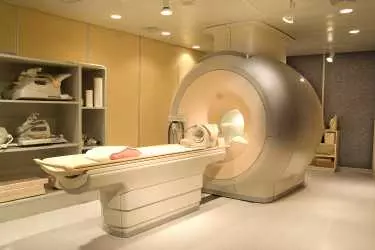
The technologist begins by positioning you on the CT examination table, usually lying flat on your back. Straps and pillows may be used to help you maintain the correct position and to help you remain still during the exam. If contrast material is used, depending on the type of exam, it will be swallowed, injected through an intravenous line (IV) or, rarely, administered by enema. Next, the table will move quickly through the scanner to determine the correct starting position for the scans. Then, the table will move slowly through the machine as the actual CT scanning is performed. Depending on the type of CT scan, the machine may make several passes.
You may be asked to hold your breath during the scanning. Any motion, whether breathing or body movements, can lead to artefacts on the images. This loss of image quality can resemble the blurring seen on a photograph taken of a moving object. When the examination is completed, you will be asked to wait until the technologist verifies that the images are of high enough quality for accurate interpretation. The CT examination is usually completed within 30 minutes. The portion requiring intravenous contrast injection usually lasts only 10 to 30 seconds.

In many ways CT scanning works very much like other x-ray examinations. Different body parts absorb the x-rays in varying degrees. It is this crucial difference in absorption that allows the body parts to be distinguished from one another on an x-ray film or CT electronic image. In a conventional x-ray exam, a small amount of radiation is aimed at and passes through the part of the body being examined, recording an image on a special electronic image recording plate. Bones appear white on the x-ray; soft tissue, such as organs like the heart or liver, shows up in shades of gray, and air appears black.
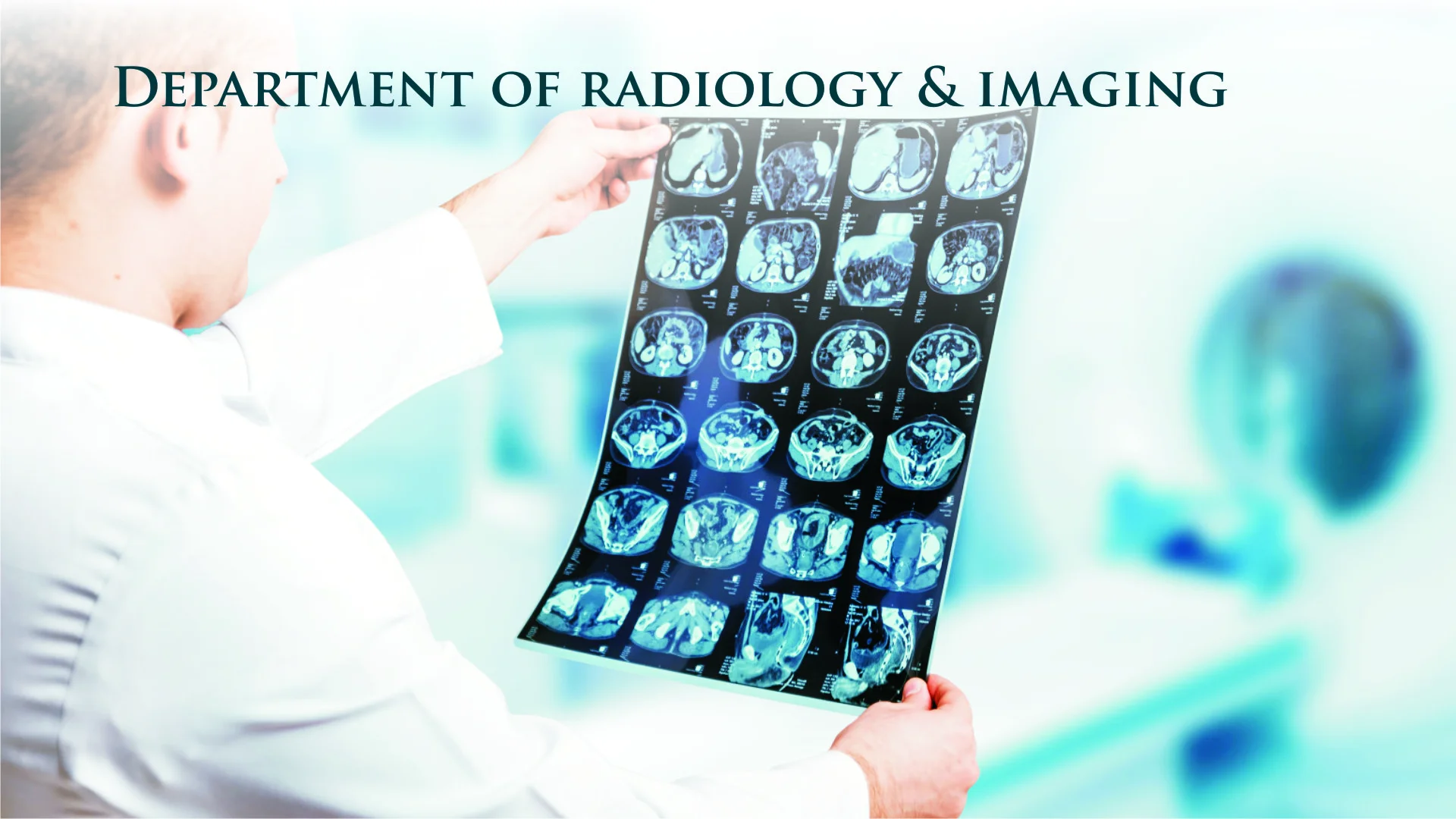
With CT scanning, numerous x-ray beams and a set of electronic x-ray detectors rotate around you, measuring the amount of radiation being absorbed throughout your body. Sometimes, the examination table will move during the scan, so that the x-ray beam follows a spiral path. A special computer program processes this large volume of data to create two-dimensional cross-sectional images of your body, which are then displayed on a monitor. CT imaging is sometimes compared to looking into a loaf of bread by cutting the loaf into thin slices. When the image slices are reassembled by computer software, the result is a very detailed multidimensional view of the body's interior.
Refinements in detector technology allow nearly all CT scanners to obtain multiple slices in a single rotation. These scanners, called multislice CT or multidetector CT, allow thinner slices to be obtained in a shorter period of time, resulting in more detail and additional view capabilities. Modern CT scanners are so fast that they can scan through large sections of the body in just a few seconds, and even faster in small children. Such speed is beneficial for all patients but especially children, the elderly and critically ill, all of whom may have difficulty in remaining still, even for the brief time necessary to obtain images. For children, the CT scanner technique will be adjusted to their size and the area of interest to reduce the radiation dose. For some CT exams, a contrast material is used to enhance visibility in the area of the body being studied.
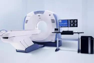
At Siddh Diagnostic Centre in Gurgaon we have the state of the art most advanced CT scan from GE Healthcare, USA. CT Scan is a great diagnostic modality for several body parts but it has to be ordered judiciously because of the Radiation dose given to the patient. . At Siddh Diagnostc Centre in Gurgaon we have CT scan which uses ASIR technology to reduce the radiation to 60% lesser than any of the CT Scan machines available in Delhi and Gurgaon. Till now the practice was to put more radiation inside the patient in order to achieve good quality images. ASIR technology has made this redundant which is of great advantage to the patient. Each CT Scan would give a patient radiation equivalent to approximately 70 X-Rays but with ACIR technology this has come down to almost 20 X-Rays.

![]() NCCT Brain
NCCT Brain
![]() CECT Brain
CECT Brain
![]() CT PNS - Axial & Coronal
CT PNS - Axial & Coronal
![]() CT Neck
CT Neck
![]() HRCT Chest
HRCT Chest
![]() CT Abdomen
CT Abdomen
![]() CT KUB
CT KUB
![]() CT Angiography
CT Angiography
![]() Whole Body CT Screening
Whole Body CT Screening

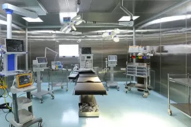
Siddh Hospital has got cutting edge advanced medical technology to take care of most complex of the diseases. Latest Flat Panel Cath Lab from Siemens, Germany is capable of doing the most difficult Coronary Procedures. The Radiology department has 64 Slice CT Scanner, 4D Ultrasound and Colour Doppler, Digital X-Ray, Mammography, DEXA Scan while the Cardiology Department has the facilities of Echocardiography, Treadmill Test, Holter and ECG. The Hospital has fully modular operation theatres and fully equipped ICU and CCU. Siddh Hospital has inhouse blood bank with component seperator and a dialysis centre.

One of the biggest obstacles to Universal Healthcare Access is the high cost of tertiary care in India. A lot of people cannot afford this costly treatment which is either not available in the public sector or there is a long waiting. Siddh Hospital brings to you the latest cutting edge healthcare delivered by the finest of the doctors at a very affordable cost. Our packages are fixed unlike many other hospitals where cost overruns are to be paid by the patient. The packages at Siddh Hospital are fixed for the patient regardless of the length of stay due to any kind of complications arising during the treatment process.
Our Chief Cardiologist Dr Anurag Mehrotra is talking about the Heart Disease, its causes and how to prevent it. Dr Mehrotra is also talking about the treatments available for Heart Ailments.
Take a Virtual Tour of Siddh Hospital in this video to know what all services are being offered by the hospital and the state of art modern technology deployed for providing world class treatment.
